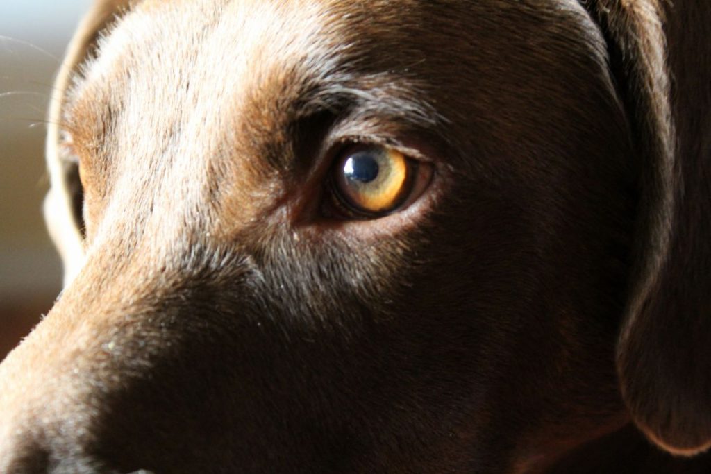
What is Uveitis in Dogs?
Inflammation of one or more structures in the eye’s uveal tract causes uveitis in dogs. The uvea is the vascular part of the eye and consists of three structures, the iris, the ciliary body, and the choroid.
Panuveitis includes all three parts of the uvea. Anterior uveitis in dogs includes the iris and the ciliary body, and posterior uveitis only involves the choroid.

What Causes Canine Uveitis?
Understanding the eye’s anatomy helps explore the causes of inflammation in the uveal tract.
The three chambers of the eye include:
- The anterior chamber consists of the space between the cornea and the iris.
- The posterior chamber consists of the space between the iris and the lens.
- The posterior segment consists of the space between the retina and the lens.
The three chambers have layers which include the sclera, the choroid, and the retina. These layers help maintain the shape of the eye, produce vitreous humor and collect light on the rods and cones to transport to the brain to facilitate vision, respectively.
The uvea’s functions include producing aqueous humor and forming the blood-eye barrier. The aqueous humor fluid produced by the ciliary body fills the anterior chamber and maintains the eye’s shape. The fluid circulates through the eye and drains through the drainage angle. The muscles around the uvea regulate constriction and dilation of the pupil.
The uvea maintains intraocular pressure and supports the lens. The essential nutrients produced by the ciliary body help support the eye’s functions and efficiency.
The uveal tract is a sensitive part of the eye. A breakdown in the eye’s blood supply barrier results in white blood cells and other inflammatory proteins entering the aqueous humor. This inflammation causes the eye to become hazy and can also change the color of the iris.
Dog uveitis causes include internal and external factors that lead to the disruption of the blood-eye barrier. An in-depth discussion of the etiology of uveitis follows below.
Etiology of Uveitis in Dogs
Establishing the etiology of uveitis requires a systematic clinical evaluation, complete medical history, and several diagnostic tests. Most evaluations include full blood counts, serum biochemistry, and infectious disease testing if applicable to the patients’ history and geographic exposure risks.
The long list of possible causes of uveitis highlights the importance of a thorough physical eye examination.
The table below lists the possible infectious and noninfectious causes of uveitis in dogs.
| Causes of Uveitis in Dogs | |
| Infectious | Non-Infectious |
| Bacterial – Borrelia burgdorferi (Lyme disease). – Brucella canis. – Ehrlichia canis. – Leptospira species. – Rickettsia rickettsii (Rocky Mountain spotted fever). Fungal – Aspergillus species. – Blastomyces species. – Coccidioides species. – Cryptococcus species. – Histoplasma species. Viral – Adenovirus (canine infectious hepatitis). – Distemper virus. – Herpes virus. – Rabies virus. Parasitic – Dirofilaria immitis. – Leishmania species. – Toxoplasma species. Other – Prototheca species. – Septicemia. |
Systemic disease – Cataracts – can cause immune-mediated uveitis when the immune system reacts to lens proteins leaking out of its capsule. – Coagulopathies. – Diabetes mellitus. – Hyperlipidemia. – Hypertension. – Lens-induced uveitis in dogs is also common. Immune dysfunction – Immune-mediated or idiopathic uveitis. – Ulcerative keratitis. – Uveodermatologic syndrome. Genetic – Pigmentary uveitis in golden retrievers. Neoplasia – Primary neoplastic disease (ocular melanoma). – Secondary neoplastic disease. Trauma |
Some studies indicate that 25 % of uveitis cases occur due to metastatic neoplasia, 17 % occur due to infectious causes, and the remaining 58% of dogs receive a diagnosis of immune-mediated or idiopathic uveitis.
Clinical Signs of Uveitis in Dogs
Most owners notice that their pets’ eyes become severely reddened, cloudy, and excessively teary. Uveitis induces extremely painful symptoms, most often exhibited through blepharospasm (shutting of the eye) and photophobia in dogs.
Listed below are explanations of ophthalmic terms and specific signs a vet examines the eye for when investigating a possible uveitis case:
- Episcleral erythema, which is the reddening of the border of the white part of the eye – the sclera. The redding occurs due to enlarged blood vessels in dogs’ eyes.
- Third eyelid elevation due to enophthalmos caused by decreased intraocular pressure and pain.
- Self excoriation marks are due to rubbing at the eye with paws or rubbing the face on furniture or carpets.
- Photophobia occurs due to the pain a dog experiences in bright lights as a result of uveitis.
- Epiphora is a result of increased tear production to “wash out” the eye.
- Decreased vision or blindness.
- Corneal edema occurs when the cornea becomes more permeable due to inflammatory mediators. The increased permeability results in a cloudy or blue appearance of the cornea.
- Dense peripheral corneal neovascularization describes new blood vessels forming at the edges of the cornea.
- Aqueous flare occurs when a small, direct beam of light creates a “headlights in fog” effect in the eye’s anterior chamber. The Tyndall effect is another term for aqueous flare.
- Hypopyon describes puss in the anterior chamber.
- Hyphema describes blood in the anterior chamber.
- Iris hyperemia or “rubeosis irides” occurs when the iris becomes reddened due to increased blood flow.
- Iris swelling, also known as iris bombe, occurs when the iris bulges outwards.
- Iris color change (obviously visible in patients with lighter irides).
- Anterior synechiae dog symptoms include the iris adhering to the cornea, and posterior synechiae dog symptoms include the iris adhering to the lens.
- Miosis and resistance to pharmacologic dilation help to differentiate uveitis from conjunctivitis.
- Low intraocular pressure occurs due to an increased outflow of fluid from the eye resulting in ocular hypotension. Measurements of <5 mmHg indicate low pressure in dogs’ eyes and support a uveitis diagnosis.
In most cases, the degree of ocular pain correlates with the number and severity of ophthalmic findings. However, this is not the case in patients with posterior uveitis or neoplasia-induced uveitis. These patients will only have severe pain if they develop concurrent glaucoma secondary to cancer.
Patients are at risk of developing secondary glaucoma with acute or chronic uveitis, leading to possible irreversible vision impairment or loss.
Chronic uveitis cases have the potential to progress into cataracts, blindness, or lens luxation/displacement.
How is Canine Uveitis Diagnosed?
The first steps in diagnosing uveitis require a full eye exam.
The eye exam will include the following steps:
- Direct ophthalmoscopy.
- Indirect ophthalmoscopy.
- Pupillary light reflex.
- Tonometry.
- Slit-lamp examination.
- Topical application of epinephrine or phenylephrine drops to the eye to differentiate between episcleral erythema and conjunctivitis. The medication causes constriction of the superficial blood vessels of the conjunctiva but not the deeper vasculature of the episclera.
An eye exam reveals pathognomonic signs of uveitis, such as aqueous flare coupled with decreased intraocular pressure and constricted pupils.
By combining clinical findings and patient history, a clinician determines which diagnostic tests are necessary to establish the inciting cause of uveitis. Each patient will present with a variety of symptoms which can be overwhelming, but it is important to recognize uveitis symptoms and signs rapidly to avoid long-term complications.
Secondary ocular changes due to uveitis can occur quickly or over a long period if the patient suffers from a recurrent or unresolved primary disease.
Some chronic changes that indicate previous uveitis events include:
- Iris hyperpigmentation, whereby pigment deposits on the anterior lens capsule. Sometimes referred to as footprints of synechia.
- Chorioretinal scars become visible well-defined hyperreflective lesions in the tapetal fundus. They present as depigmented lesions in the non-tapetal fundus.
Glaucoma and uveitis have several similar clinical signs but there is a specific diagnostic test used to determine which condition is present. Clinicians use tonometry to measure intraocular pressure. Uveitis decreases intraocular pressure, whereas glaucoma results in increased intraocular pressure.
Ultrasound examination of the eye offers a unique diagnostic tool to visualize the chambers and components of the inner eye in a non-invasive manner. In the case of neoplasia, CT imaging referrals occur if a lesion is rooted deep within the ocular tissue or orbit.
Determining the primary cause of uveitis requires the skills of multiple veterinary disciplines, including internal medicine, ophthalmology, radiology, and possibly infectious disease specialists. Due to the high costs of diagnosing and treating uveitis in a patient, owners need to prepare themselves financially.

Uveitis in Dogs – Treatment Options
The most important part of treatment for uveitis aims to stabilize the blood-eye barrier, maintain vision, reduce inflammation in the eye, and provide pain relief for the patient.
Topical eye medications containing corticosteroids, non-steroidal anti-inflammatory drugs, or mydriatic agents provide some relief, but often patients require a multimodal approach for pain relief.
Examples of topical corticosteroids include prednisolone or dexamethasone. Clinicians avoid prescribing hydrocortisone. Hydrocortisone does not penetrate the cornea effectively, preventing it from achieving therapeutic levels that facilitate pain relief.
Common topical NSAIDs include flurbiprofen sodium, diclofenac sodium, and ketorolac tromethamine.
The most common mydriatic and cycloplegic medication (causes pupil dilation and ciliary muscle relaxation, respectively) is topical atropine ointment or solution. The atropine relieves ciliary spasms, provides comfort, and helps to stabilize the blood-eye barrier. Use mydriatic medications with caution because they can exacerbate glaucoma.
In cases of uveitis caused by trauma, referral specialists often perform surgery to repair corneal tears or remove foreign bodies.
The best course of treatment always depends on diagnosing the underlying cause in the case of non-traumatic uveitis. Infectious causes may need antibiotics, antiviral eye drops, or antifungal medications.
If topical medications prove ineffective, clinicians turn to systemic NSAIDs or corticosteroids to attempt to control uveitis. Systemic corticosteroids pose a risk in infectious causes of uveitis as they may suppress the immune system and exacerbate the infection.
Immune-mediated cases sometimes call for immunosuppressive drugs to control uveitis. When treating uveitis, azathioprine or mycophenolate are options, but patients need careful monitoring to avoid decreased white blood cell, red blood cell, and platelet counts.
A natural treatment for uveitis in dogs investigated by scholars at the Texas A&M College of Pharmacy shows good potential in managing uveitis.
The team discovered the anti-inflammatory properties of curcumin, a compound found in turmeric. When processed into a unique nanoparticle formulation, it showed positive findings to aid in managing uveitis in humans and animals.
Uveitis resolution is a long road, and some owners discontinue medication before the underlying cause is effectively addressed or successfully treated. The frustrating idiopathic uveitis or immune-mediated uveitis cases require chronic medication and a lifelong commitment to daily medication administration to pets.
Pathology of Canine Uveitis
Uveitis can be unilateral or bilateral, and it presents in a significant number of ophthalmology cases.
The pathophysiology of uveitis begins after the uveal tissue or vasculature undergoes damage or if anything disrupts the ocular blood-eye barrier. The blood-eye barrier consists of an epithelial and an endothelial layer. Tight junctions form between the retinal capillaries and the retinal pigment epithelium cells.
The barrier protects the eye by preventing molecule movement across the vascular endothelial surface and maintains the protein concentration of the aqueous humor.
If the barrier undergoes any damage, the aqueous humor protein concentration increases, resulting in light scattering. This sign, known as the Tyndall Effect, illuminates a slit beam within the anterior chamber.
The resultant aqueous flare is a hallmark of uveitis. The blood-retinal barrier disruption leads to retinal edema, retinal hemorrhage, and possible detachment of the neurosensory retina.
In the acute inflammatory phase of uveitis, arteriolar vasoconstriction occurs briefly, and prolonged vascular dilation follows. The mediation of vasodilation occurs due to prostaglandins and leukotrienes that cause increased vascular permeability. The increased vascular permeability results in the breakdown of the blood-eye barrier.
Prostaglandins induce hyperemia, reduce intraocular pressure, and constrict the iris sphincter muscle, which then causes miosis and pain.
As the blood-eye barrier breaks down, proteins, cells, and additional inflammatory mediators enter the iridal stroma and aqueous humor. These processes, in turn, lead to a decrease in intraocular pressure.
The Zoonotic Potential of Canine Uveitis
Several infectious diseases have the potential to cause uveitis in both humans and animals. Zoonotic transmission of diseases can occur between pets and their owners or between patients and their attending veterinary staff. If a patient presents with uveitis – staff must take necessary precautions and work with protective clothing.
Leptospirosis
Uveitis due to Leptospirosis occurs in humans, dogs, and horses. Leptospirosis features as one of the most common global zoonoses. Dogs infected with leptospirosis sometimes develop uveitis before developing any other clinical signs.
Typical signs of Leptospirosis include lethargy, depression, loss of appetite, vomiting, fever, icterus, bleeding diathesis, and increased thirst and urination. The illness develops quickly with clinical outcomes ranging from mild morbidity to abrupt fatality.
Some animals only develop subclinical manifestations of the disease and remain undetected.
A large number of serovars for Leptospirosis that cause uveitis means that even though vaccines are available, not all of the strains form part of the vaccines. Clinicians test any animal presenting with uveitis if they originate from an area with endemic Leptospirosis.
The transmission of leptospirosis bacteria occurs through contact with infected urine.
Brucellosis
Brucella canis causes uveitis in both humans and dogs. All intact dogs with a breeding history presenting to a clinic with uveitis carry a risk of Brucellosis infection to veterinary staff.
Male dogs infected with brucellosis develop epididymitis, scrotal swelling or enlarged testicles, and scrotum skin rash. The dog may be infertile. In chronic or long-standing cases, the testicles will atrophy or become shrunken.
Female dogs infected with brucellosis develop pyometra and infertility and often abort in the late stages of pregnancy. Lymphadenopathy and systemic infection result in more severe symptoms.
All staff must exercise caution and wear protective clothing when working with suspected Brucellosis cases. Staff must take great care if members are pregnant or trying to conceive. There is no vaccination against Brucellosis, treatment takes a very long time, and severe illness sometimes results in permanent infertility.
The Prognosis of Canine Uveitis
Treatment for most cases of uveitis provides relief for most patients within 24 hours. Addressing the underlying cause of the uveitis is the primary step in avoiding recurrence or incomplete resolution.
The more severe the clinical symptoms, the longer the patient will take to recover. Complications and chronic cases may lead to irreparable and permanent vision impairment or loss. Chronic canine uveitis and cancer complications carry a guarded prognosis.
Some complications can include:
- Cataract formation (usually capsular and outer cortex).
- Iris bombé due to complete circumferential posterior synechiae.
- Lens subluxation or luxation.
- Phthisis bulbi.
- Pre-iridal fibrovascular membrane.
- Retinal degeneration.
Is Uveitis in Dogs Contagious to Humans?
The two conditions transmissible to humans that have the potential to cause uveitis include Leptospirosis and Brucellosis.
Leptospirosis infection occurs when humans come into contact with infected animal urine. Dogs can carry urine on their fur or paw pads and act as fomites for the bacteria. Pet owners or people who regularly handle dogs or clean kennels are at risk of contracting Leptospirosis.
Personal protection and high standards of personal hygiene are the most effective means to avoid zoonotic infections in high-risk environments.
Brucella canis poses a zoonotic risk to veterinary personnel, especially for women who are pregnant or trying to conceive. Brucellosis can cause spontaneous abortion as well as uveitis. Breeding dogs with systemic, infectious uveitis should undergo testing for Brucellosis. There is no vaccine for Brucellosis canis, so working cautiously with high-risk patients is important.

The Final Word
The subtle symptoms and complex sequelae of uveitis make it a frustrating condition. The condition presents as a complicated diagnosis for owners. Veterinary staff need to be able to convey the condition in simple and concise terms without overwhelming clients. Owner compliance is a critical step in managing to treat a patient’s condition successfully.
Determining the underlying cause of uveitis is key for treatment and avoiding potential zoonotic risks for both owners and veterinary personnel. Uveitis patients suffer from severe pain, and owners need to commit to long-term treatment for their pets. Commitment to treatment offers freedom from discomfort with the aim of preserving vision.

