
A biopsy is one of the most important diagnostic tools in veterinary medicine. It’s so important that I can’t think of a veterinarian who never does a biopsy. Whether it’s a dermatologist, dentist, surgeon, internal medicine specialist or your primary care veterinarian, all use biopsies to diagnose different conditions.
At the Schwarzman Animal Medical Center, veterinarians submit between five and fifteen biopsies on any given day. In many cases, a biopsy is the only way to confirm a diagnosis, especially for internal diseases like inflammatory bowel disease, liver problems and most kinds of cancer.
In today’s blogpost, I’ll discuss how biopsies work and why they’re essential for diagnosis.
What is a biopsy?
A biopsy is a diagnostic procedure where a piece of tissue is taken from the body for examination. Depending on where the tissue is located, the sample could be obtained using a skin punch biopsy instrument, via ultrasound guidance, with endoscopic equipment or during a surgical procedure. Once obtained, the tissue sample is then processed in a laboratory to preserve the cells and their arrangement in the tissue. The sample is then sliced so thin that light can pass through it, and it’s stained to highlight different cells. This allows the tissue to be examined using a microscope by a veterinary pathologist.
What is in a biopsy report?
Like any report, a biopsy report starts with the who, what and where:
Who is the patient’s name and owner’s name
What indicates dog, cat or other species, and
Where is the location of the sample.
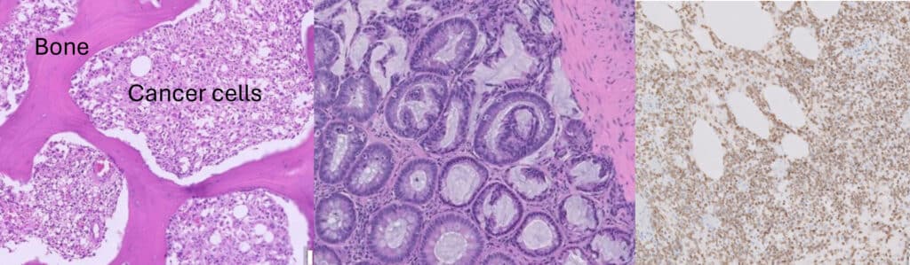
Next, the pathologist describes what they are seeing. There are usually cells of some type, perhaps round or spindle shaped. For example, in Panel 1 of the image below, the purple and white blobs are dozens of cancer cells called histiocytic sarcoma. The bright pink material surrounding the cancer cells is bone.
How do veterinarians use biopsy reports?
Biopsy reports often contain very detailed information that helps clinical veterinarians make treatment recommendations and prognostic predictions. For example, liver specialists look at copper levels in the liver to help them treat hepatitis. Oncologists focus on the number of dividing cells in the biopsy to predict cancer outcomes. Finally, the biopsy report will give a diagnosis, such a copper associated liver disease or an ear polyp in a young cat.
In Panel 2 above, this biopsy from a cat indicates inflammatory bowel disease.
When does a biopsy not give a diagnosis?
There are occasions when a pathologist cannot make a diagnosis from a biopsy. Sometimes two diagnoses can have a similar appearance, and the pathologist needs more information. At other times, the cells are cancerous, but so abnormal that the pathologist cannot determine what type of cancer is present—only that the diagnosis is cancer. In these cases, pathologists use a special technique called immunohistochemistry to better identify the type of cells present in the biopsy. From that information, they can often make a diagnosis. Immunohistochemistry takes time and the wait is hard for pet owners and their veterinarians as well. Panel 3 shows an example of an immunohistochemical stain used to diagnose intestinal lymphoma in a dog.
To learn more about how a biopsy sample is processed, read my article “Biography of a Biopsy,” published on Vet Street several years ago.